Children with Osteogenesis Imperfecta
The term osteogenesis imperfecta (OI) began with William Vrolik in the 1840s. OI is often known as brittle bone disease, and is a genetic connective tissue disorder, involving type 1 collagen – An issue with the quality of type I collagen may cause a more moderate or severe form of OI, whereas an issue with the quantity of type I collagen may cause more mild forms of OI (1)
OI is characterized by:
- low bone density
- fractures
- spine and extremity deformity
and clinical presentations are variable.
The better understanding of the genetics of OI, has seen considerable addition to the Sillence classification of basic OI types, especially in the last decade, and now includes a non-type 1 collagen OI type:
Sillence OI classification (4 types)
Type I: Mild, nondeforming
Type II: Lethal perinatal
Type III: Severe, progressively deforming
Type IV: Phenotypically variable with white sclera
[Type 5 – not associated with type 1 collagen]In the last 10 years, many more variations of OI have been identified. These approximate 15% of OI, due to genetic mutation affecting the synthesis of type I collagen. If each specific genetic cause is a type, there are now some 20 OI types. This is important to be aware of, but clinically the ‘Sillence + model’ is still helpful (+ represents the increasing array of genetic typing). A new nomenclature for OI has combined causative genes, phenotypes and severity ranges (2).
Clinical presentations of OI vary
Just as the role of type 1 collagen is varied, so too are the range and severity of the skeletal manifestations of OI.
| Common skeletal inclusions | Non-skeletal inclusions |
|---|---|
| · low bone mass | · blue sclerae |
| · history of fractures | · hydrocephalus |
| · bowing of the long bones (legs/arms) | · hearing loss |
| · vertebral compression fractures | · dental dystrophy |
| · basilar invagination | ·
|
| · scoliosis | |
| · spondylolisthesis | |
| · ligamentous laxity | |
| · joint deformities | |
| · short stature |
Multi-disciplinary medical and health care is beneficial. The principles of managing OI focus on nutrition and physical activity, with adequate calcium and vitamin D being essential for bone health. Bisphosphonates as antiresorptive drugs may be used to improve bone mass and vertebral structures, and hopefully reduce fracture frequency. However studies are variable in protocols, design, and overall quality which makes clinical decisions less certain(3).
Physical activity is really important to reduce skeletal deformity and fracture risk, and needs to suit each child’s varying needs, and age. The relationship between muscle strength and bone strength is really important for children with OI.
The aims of health care for children with OI depend very much of age and needs, but always promote independence and in pre-schoolers the focus is on achieving milestones as near to same-age peers as possible.
Common gait deviations with OI and the use of foot orthoses:
There is much variation, but common gait pattern changes and features are:
- increased external hip rotation
- decreased strength of the hip flexors
- decreased strength of abductors
- decreased toe-off propulsive strength.
Exercises for improving core and hip strengthening can help both gait and posture.
Children with OI may have flatfeet that disadvantage gait or cause pain. Not all children with OI and flatfeet require orthotics, and every child with OI needs to be individually assessed. In my experience, strength and footwear selection is paramount, and the use of orthoses reserved for those who need help with gait and activity, or foot pain. Outcome measures are helpful to assess intervention effects, and the PODCI has been found reliable in OI management (4).
The p-GALS gait assessment is a really helpful approach generally, and given the increased likelihood of scoliosis with OI, p-GALS is especially relevant (5). The prevalence of scoliosis with OI is cited as 39% to 80%, rarely seen before 5-6 years, but can progress rapidly.
Case example
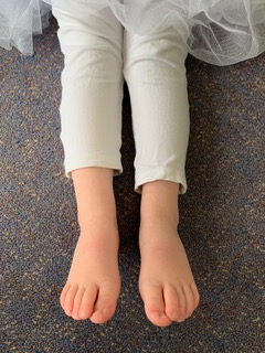
A 3-year-old child with OI (familial, type 1*; known across four generations) referred with orthopaedic and physiotherapy concerns about tib/fib stress due to child’s genu valgum/flatfeet and gait instability, with more than usual falls/age. Ingrown nail tendency was also noted, with no history of infections.
This child was otherwise well, with generalised hypermobility (Beighton score 7/9).
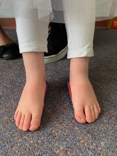
The current footwear was rigid-sole boots, which aimed to stablise the feet, but actually reduced effective toe-off, and possibly promoted tripping.
Genu valgum was increased in stance, and gait unstable due to joint hypermobility, and pronated feet. This little girl would fall when bending over to pick up a leaf (etc..), and could not run safely, given her increased fracture risk.
In addition to continuing physiotherapy and strength-based activity, footwear was changed to a lighter weight runner (Asics Contend), and supplemented with shortened and posted EVA heel cups, to stabilise foot posture, and better align knees.

Three months later, she could squat (vid 1) and run (vid 2). She had progressed to kicking a ball, and knee x-rays demonstrated normal alignment without weight-bearing. Whilst ingrown nail tendency persists, the parents reported that there was generally less redness.
* Non-Deforming OI With Blue Sclerae—OI Type 1 (2):
- OI type 1 is characterized by increased bone fragility, which is usually associated with low bone mass, distinctly blue-grey sclerae, and susceptibility to conductive hearing loss, beginning in adolescence.
- Deformity of long bones or spine is uncommon, and where scoliosis develops, it is commonly an idiopathic.
- OI type 1 is the most common variety of OI in European derived communities and has a birth prevalence in the order of 1:25,000 live births and a similar population frequency.
- Fracture frequency and mild long bone and spine deformity mean that it is generally perceived to be of mild severity but can be moderate -severe, particularly when DI is present.
The ‘hallmark’ ocular blue sclera in:
- child of case example b. her father


Health related quality of life (HRQoL) with OI:
A recent case-control study (n=128) found that children with OI had significantly reduced HRQoL, with inverse correlation between OI severity and HRQoL (6).
Thank you for viewing this 2020 Evidence Essentials blog.
Kind regards,
Angela Evans
Dr Angela Evans AM
PhD, FFPM RCPS(Glasg)